Indications
Cardiac Testing
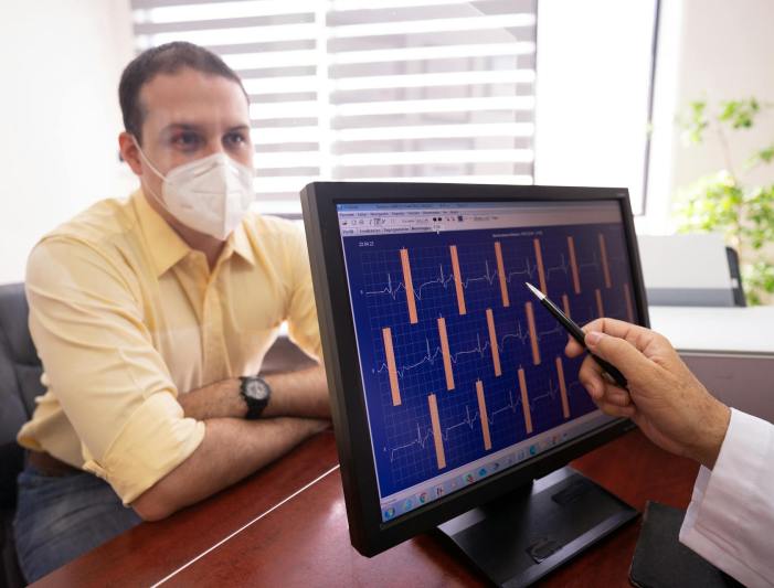
Electrocardiogram (ECG)
A non-invasive, painless test that records the heart’s electrical activity to assess rhythm and function. Small patches called electrodes are placed on the chest, arms, and legs. These electrodes connect to a machine that traces the electrical signals, creating a visual record on paper. This tracing allows doctors to diagnose various heart conditions, including arrhythmias (irregular heartbeats), blocked arteries, and heart defects.
Exercise Stress Test (GXT)
Also know as a cardiac stress test. A diagnostic procedure that evaluates your heart’s function during physical exertion. You typically walk on a treadmill or ride a stationary bike to increase your heart rate. Throughout the test, a healthcare professional monitors your heart rate, blood pressure, and continuous electrocardiogram (ECG) readings to see how your heart responds to the increased demand.
2D Doppler ECHO
A non-invasive ultrasound that uses high-frequency sound waves (Doppler Technology) to produce two-dimensional images of the heart’s structure and function. Analysis of the speed and direction of blood flow through the heart can help detect blockages, valve leakage, congenital heart disease, and cardiomyopathy.
Stress Test with Cardiac Enhancer
A heart test for patients who cannot perform physical exercise (like walking on a treadmill). An IV medication called dobutamine is administered to increase the heart rate, simulating the effects of physical stress. An echocardiogram is then allowing doctors to evaluate and assess blood flow and the structure and function of the heart valves while under stress.
Speckle Tracking ECHO
An echocardiographic technique that measures myocardial deformation by tracking the motion of natural speckle patterns within the heart muscle. It measures strain providing an assessment of heart function that can reveal congenital defects and other issues. This information helps diagnose and monitor heart conditions and can be used for risk assessment and determining the optimal time for intervention.
Cardiac Event Loop Recorder
A small, implantable device that continuously monitors the heart’s rhythm to help diagnose heart conditions that cause symptoms like fainting, palpitations, or dizziness. It is placed under the skin and can record for up to three years, capturing abnormal heart rhythms that may not be detected by routine tests. Can be programmed to record automatically or to be activated by the patient when symptoms occur.
Bubble Study Echocardiogram
A bubble study (or bubble echo) is a heart ultrasound that uses an injection of tiny, agitated saline bubbles. By watching how these bubbles travel through the heart, doctors can check for abnormalities like a hole in the heart and help find the cause of conditions like unexplained strokes.
Holter Monitor
A type of portable cardiac monitor for electrocardiography that records the electrical activity of the heart. Worn by the patient continuously for periods ranging from 48 hours to 30 days.
Ambulatory Blood Pressure Monitor
A test that uses a small, portable blood pressure monitor worn by the patient. Measures and records blood pressure at regular intervals throughout the day and night during normal daily activities and while sleeping, providing a detailed profile that helps diagnose hypertension and related issues.